
Grade 1, 2 and 3 describe one thing: how much fat is scattering the ultrasound beam as it passes through your liver. None of the three grades measures scarring, and scarring is what decides outcome. All-cause mortality runs 0.32 per 100 person-years at fibrosis stages 0–2 against 1.76 per 100 person-years at cirrhosis (AASLD Practice Guidance, 2023).
This article is for people in Chennai who have just read one line at the bottom of an abdominal ultrasound report — grade 1 fatty liver, or grade 2, or fatty infiltration of the liver, usually before any doctor has explained it. It covers what the grade describes, how reliable that grade is, which tests come next, and what the published evidence says actually changes the liver. It is not about hepatitis testing pathways, and it is not a substitute for reading the whole report with the doctor who ordered it.
One boundary first, because the anxiety this finding causes sends people to the wrong place. A fatty liver reported on a routine scan is not an emergency and does not need same-day review. But yellowing of the eyes or skin, swelling of the abdomen, vomiting blood, black tarry stools, or new confusion or drowsiness are hospital problems, not outpatient ones. AJSMC in Egmore, Chennai is an outpatient and day-care centre with no emergency department and no critical care — its ten beds are for planned, stable admissions, with a nurse on site overnight and no doctor on the premises — and it is closed outside Monday to Saturday, 10am to 9pm. For any of those signs, at any hour, go to a hospital with a 24-hour emergency department or call 108.
What does grade 1, 2 or 3 fatty liver actually mean?
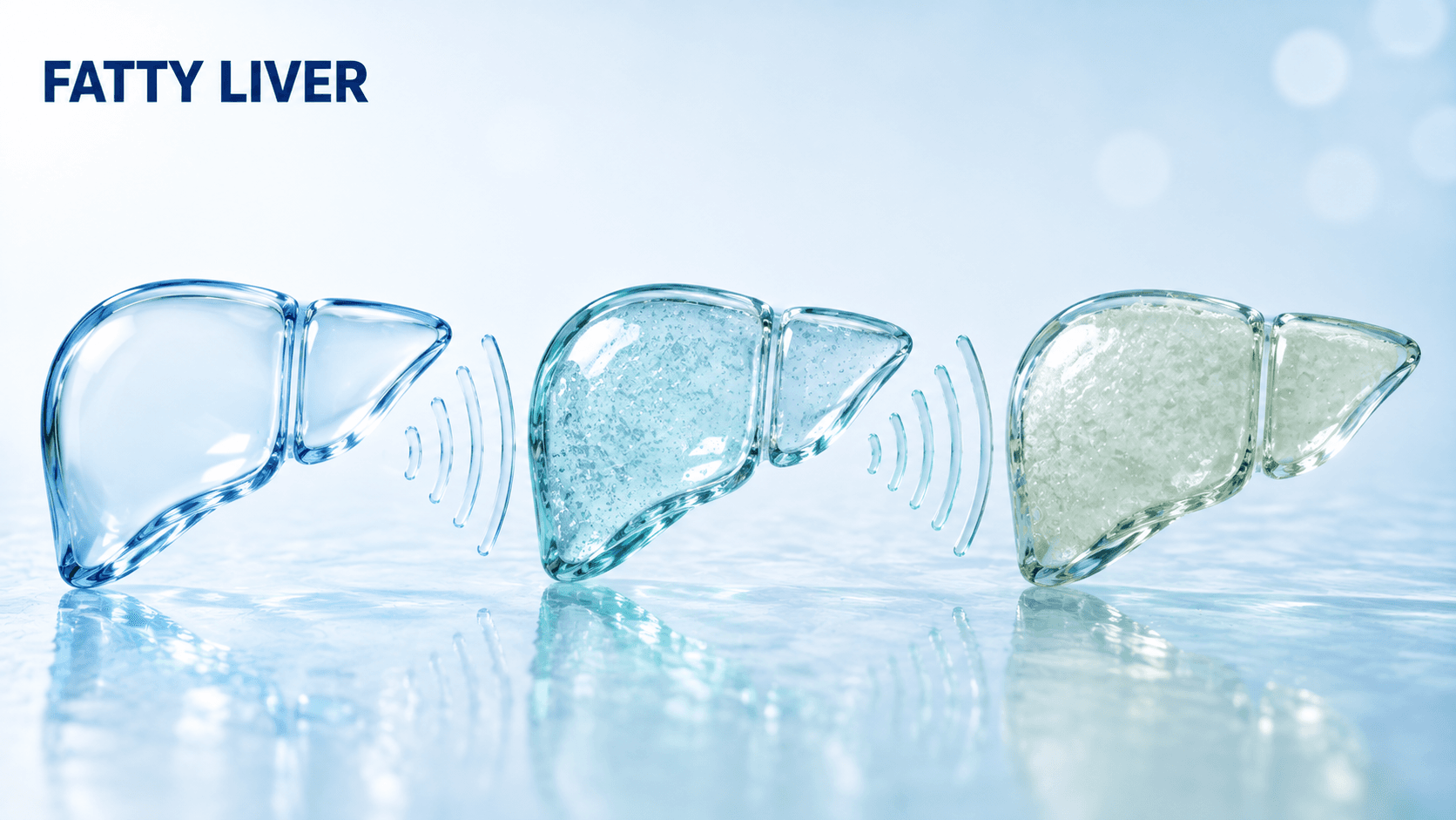
It means the radiologist judged three things by eye: whether the liver looks brighter than the kidney beside it, whether the walls of the portal veins and the diaphragm are still visible through it, and how far the ultrasound beam still penetrates (Korean Journal of Radiology, 2022). The grade is a description of the picture, not a stage of disease.
| Grade | What the radiologist saw | What it does not tell you |
|---|---|---|
| Grade 1, mild | Liver brighter than the kidney; diaphragm and portal vein walls still clearly seen | Nothing about scarring, inflammation or prognosis |
| Grade 2, moderate | Liver brighter still; the diaphragm and portal vein walls becoming blurred | Nothing about scarring, inflammation or prognosis |
| Grade 3, severe | Liver markedly and unevenly bright; the beam no longer penetrates; diaphragm obscured | Nothing about scarring, inflammation or prognosis |
Source: Korean Journal of Radiology, 2022.
Two consequences follow, and they are the reason this page exists. A grade 1 liver can carry advanced scarring, and a grade 3 liver can carry none. So a patient in Chennai who has been told "only grade 1, nothing to worry about" and a patient told "grade 3, this is serious" have both been given information the scan cannot supply.
How reliable is the grade itself?
Less reliable than the confident single line on the report suggests. Agreement between two radiologists grading the same B-mode ultrasound is moderate at best, and the same reader grading the same liver twice does not always return the same answer.
| Measure | Value | What it means in practice |
|---|---|---|
| Agreement between two observers | kappa 0.43 | Two radiologists frequently disagree on the grade |
| Agreement of one observer with themselves | kappa 0.54 | The same reader can grade the same liver differently |
| Sensitivity for mild steatosis (over 0–5% fat) | 73.3% | Roughly one in four mild cases is missed |
| Accuracy in people with obesity | Reduced | Reduced in exactly the group most likely to be scanned |
| Telling fat from scarring | Not possible on B-mode | Steatosis can have echo characteristics similar to advanced fibrosis (AASLD, 2023) |
Source: Korean Journal of Radiology, 2022; AASLD Practice Guidance, Hepatology, 2023.
The American guidance goes further than most patients expect. AASLD's Guidance Statement 17 (2023) says standard ultrasound is not recommended as a tool to identify fatty liver, because of low sensitivity across the whole spectrum of the disease. The corollary matters just as much for anyone in Chennai whose scan came back clean: AASLD states that the absence of detectable steatosis on ultrasound does not exclude steatohepatitis or fibrosis.
Is fatty liver serious, or is this an incidental finding?
Both, depending on a number the scan did not measure. Fatty liver is extremely common in India, a systematic review and meta-analysis put pooled prevalence at 38.6% of Indian adults and 35.4% of Indian children (Journal of Clinical and Experimental Hepatology, 2022). It is also not confined to people who look overweight. A separate global meta-analysis of 53 studies, covering 65,029 people with the condition, found lean fatty liver in 11.2% of the general population and in 25.3% of everyone with the condition (Hepatology Communications, 2020), a worldwide figure rather than an Indian one. The Indian meta-analysis itself reports a non-obese prevalence only for children, at 12.4%.
What separates the incidental finding from the serious one is fibrosis stage. AASLD (2023) states that fibrosis stage is the best predictor of long-term outcome, and that bridging fibrosis and cirrhosis carry an exponentially greater risk of liver-related illness and death than earlier stages.
| Fibrosis stage | All-cause mortality |
|---|---|
| Stages 0–2 | 0.32 per 100 person-years |
| Bridging fibrosis (F3) | 0.89 per 100 person-years |
| Cirrhosis (F4) | 1.76 per 100 person-years |
Prospective cohort of 1,773 patients, cited in AASLD Practice Guidance, Hepatology, 2023. Ultrasound grade 1, 2 or 3 does not appear anywhere in this table, because ultrasound does not measure fibrosis.
That is the turn of the whole subject. The grade tells you the liver has fat in it. The question that decides what happens over the next twenty years is how much scarring has developed, and answering it takes a blood-based score, sometimes elastography, and a clinician who has looked at the rest of the picture.
Why is it now called MASLD instead of fatty liver disease?
Because the definition changed, not only the label. A multisociety Delphi consensus of 236 panellists from 56 countries renamed non-alcoholic fatty liver disease (NAFLD) as metabolic dysfunction-associated steatotic liver disease, or MASLD, and NASH as MASH (Journal of Hepatology, 2023). Seventy-four per cent supported a change; "non-alcoholic" was judged stigmatising by 61% of respondents and "fatty" by 66%.
The substantive change is the logic of the diagnosis. NAFLD was a diagnosis of exclusion — fat in the liver of somebody who does not drink much, with other causes ruled out. MASLD is a diagnosis of inclusion: fat in the liver plus at least one cardiometabolic feature, which is why the blood tests that follow the scan are aimed at the metabolism as much as at the liver. Those features are raised body weight or waist measurement, raised blood sugar or established diabetes, raised blood pressure, raised triglycerides, and low HDL cholesterol, with thresholds set lower for Asian populations than for Western ones. Ask the consultant which cut-offs apply to you rather than reading a figure off a website; several are quoted inconsistently in secondary sources.
A second new category, MetALD, covers people who have the metabolic features and also drink meaningfully. It exists because the two do not simply add up, as the next section explains.
Which tests come next after the scan?
Blood, and mostly not liver blood. This is the initial work-up AASLD (2023) sets out after a fatty liver is found.
| Test | What it tells you | What it does not tell you |
|---|---|---|
| Hepatic panel — ALT, AST, alkaline phosphatase, bilirubin | Whether there is current liver injury; the values feed the FIB-4 score | Normal enzymes do not exclude steatohepatitis or advanced fibrosis (AASLD, 2023) |
| Complete blood count with platelets | The platelet count feeds FIB-4; a low count can hint at portal hypertension | Not a fibrosis test on its own |
| Fasting plasma glucose and HbA1c | Whether a cardiometabolic criterion is met; diabetes drives progression | Nothing about the liver directly |
| Fasting lipid profile | Triglycerides and HDL are two of the defining criteria | Nothing about fibrosis |
| Creatinine with urine albumin-to-creatinine ratio | Kidney risk travels with this diagnosis | Nothing about the liver |
| Hepatitis C serology, if not previously screened | Excludes a separate, treatable cause | — |
| If enzymes are raised: autoimmune serology, transferrin saturation, ceruloplasmin, alpha-1 antitrypsin | Excludes autoimmune hepatitis, iron overload, Wilson disease and alpha-1 antitrypsin deficiency | — |
Source: AASLD Practice Guidance, Hepatology, 2023, Table 1. The same guidance asks for the history alongside the bloods: weight history, current and recent medicines, family history of diabetes, fatty liver or cirrhosis, screening for obstructive sleep apnoea, and alcohol intake by amount, pattern and duration.
AJSMC has an in-house laboratory in Egmore, Chennai handling blood and urine samples, and its outpatient hours are Monday to Saturday, 10am to 9pm. Before travelling for any test named on this page, call 044 2532 2021 and ask which of them are run in the building and which are not, so a sample is not given in the wrong place.